Posterior Pelvis Anatomy Muscles : - There are many muscles that form the pelvic floor, including puborectalis, pubococcygeus, iliococcygeus and coccygeus.
byAdmin-
0
Posterior Pelvis Anatomy Muscles : - There are many muscles that form the pelvic floor, including puborectalis, pubococcygeus, iliococcygeus and coccygeus.. Muscular pelvic floor closure helps to relieve fascial stress. These muscles originate near the anteroinferior external surface of the bony pelvis and insert at the linea aspera. The anatomy of the pelvic floor (sometimes called the pelvic diaphragm) is complex and the terminology used varies between sources. These muscles, including the gluteus maximus and the hamstrings, extend the thigh at the hip in support of the body's weight and propulsion. ▪anterior wallis the shallowest wall and is formed by the posterior surfaces of the bodies of the pubic bones, the pubic rami, and the symphysis pubis.
These muscles, including the gluteus maximus and the hamstrings, extend the thigh at the hip in support of the body's weight and propulsion. Biceps femoris long and short head, semitendinosus, and semimembranosus. The gluteus maximus attaches from the posterior iliac crest, posterolateral sacrum, and coccyx to the gluteal tuberosity and itb. Tibialis posterior muscle (musculus tibialis posterior) tibialis posterior is the most central and deepest muscle located in the posterior aspect of the leg. Right posterolateral view of the lumbar spine.
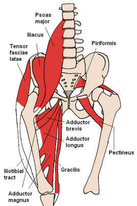
Muscles Of The Pelvis from www.learnmuscles.com Biceps femoris long and short head, semitendinosus, and semimembranosus. These muscles, including the gluteus maximus and the hamstrings, extend the thigh at the hip in support of the body's weight and propulsion. We've already identified the muscle imbalances that occur with a posterior pelvic tilt, so the solution is to fix these imbalances through either stretching and lengthening the tight muscles, and strengthening the weak ones. The anatomy of the pelvic floor (sometimes called the pelvic diaphragm) is complex and the terminology used varies between sources. Except for the short head of the biceps femoris, the other posterior thigh muscles span the length of the femur and coss both the hip and knee joints. Prominent bony landmarks are labeled. The posterior thigh is composed of three muscles: It also posteriorly tilts and contralaterally rotates the pelvis at the hip joint.
The posterior compartment is made up of a group of muscles called the hamstrings, including semitendinosus, semimembranosus and biceps femoris.
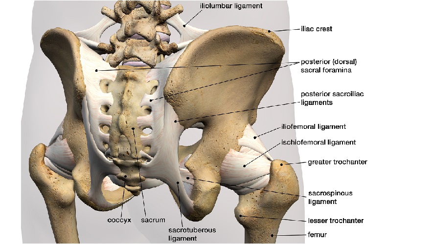
Prominent bony landmarks are labeled. Because of the lordotic curve and the thick musculature that overlies the lumbar spine, the only easily palpable bony landmark is the spinous process. ▪anterior wallis the shallowest wall and is formed by the posterior surfaces of the bodies of the pubic bones, the pubic rami, and the symphysis pubis. Posterior pelvic tilt is by far a less common postural imbalance than its opposite, excessive anterior pelvic tilt. Über 7 millionen englischsprachige bücher. They have several functions, including helping to support the pelvic organs. There are two major groups of ligaments that provide nearly all the structure of the pelvis. Muscular pelvic floor closure helps to relieve fascial stress. The pelvic floor consists of several muscles within a web of connective tissues, attaching to the bones of the pelvis, sacrum and coccyx. Arcus tendineus levator ani and the ischial spine Spanning from the posterior pelvis to the proximal tibia and fibula, the posterior thigh muscles provide motion to both the femoroacetabular joint (hip joint) and tibiofemoral joint (knee joint). Anatomy of the pelvic viscera geoffrey w. In the back of the torso, the latissimus dorsi is a large, rectangular muscle that extends from the lower back near the top of the pelvis to near the shoulder.
Because of the lordotic curve and the thick musculature that overlies the lumbar spine, the only easily palpable bony landmark is the spinous process. In the back of the torso, the latissimus dorsi is a large, rectangular muscle that extends from the lower back near the top of the pelvis to near the shoulder. The levator ani muscles are the largest group of muscles in the pelvis. These muscles originate near the anteroinferior external surface of the bony pelvis and insert at the linea aspera. The bony pelvis comes together to provide support for the pelvic muscles and connective tissues, which, in turn, provide attachments and support for the pelvic organs.
Pelvis Wikipedia from upload.wikimedia.org The three muscles within the superficial posterior compartment include the gastrocnemius, soleus, and plantaris muscles. It forms the base of the popliteal fossa and is the only muscle of either the deep posterior or superficial posterior fossa to act solely on the knee joint as a posterolateral stabilizer. These three muscles are collectively referred to as the hamstring muscles. It originates from the ischial spines and travels to the lateral aspect of the sacrum and coccyx, along the sacrospinous ligament. Right posterolateral view of the lumbar spine. Über 7 millionen englischsprachige bücher. ▪anterior wallis the shallowest wall and is formed by the posterior surfaces of the bodies of the pubic bones, the pubic rami, and the symphysis pubis. The gluteus maximus extends, laterally rotates, abducts (upper fibers), and adducts (lower fibers) the thigh at the hip joint.
The bony pelvis comes together to provide support for the pelvic muscles and connective tissues, which, in turn, provide attachments and support for the pelvic organs.
They have several functions, including helping to support the pelvic organs. Cundiff background value of surgical anatomy as in all surgical specialties, the reconstructive pelvic surgeon is frequently faced with situations that are best addressed by applying a clear understanding of the pertinent anatomy. The bony pelvis comes together to provide support for the pelvic muscles and connective tissues, which, in turn, provide attachments and support for the pelvic organs. It is situated partly within the pelvis against its posterior wall, and partly at the back of the hip joint. These muscles originate near the anteroinferior external surface of the bony pelvis and insert at the linea aspera. The popliteus is accompanied by the tibialis posterior, flexor digitorum longus, and flexor hallucis longus forming the deep posterior compartment of the leg. The levator ani muscles consist of three. It forms the base of the popliteal fossa and is the only muscle of either the deep posterior or superficial posterior fossa to act solely on the knee joint as a posterolateral stabilizer. The pubococcygeus is the intermediate part of the levator ani muscles. In this section, we'll cover the process of how to fix a posterior pelvic tilt. Therefore, an appreciation of the female pelvic musculoskeletal anatomy is critical for understanding the pelvic support system. The posterior compartment is made up of a group of muscles called the hamstrings, including semitendinosus, semimembranosus and biceps femoris. The anatomy of the pelvic floor (sometimes called the pelvic diaphragm) is complex and the terminology used varies between sources.
Right posterolateral view of the lumbar spine. Attached to the pelvis are muscles of the buttocks, the lower back, and the thighs. They have several functions, including helping to support the pelvic organs. Posterior pelvic tilt is by far a less common postural imbalance than its opposite, excessive anterior pelvic tilt. Biceps femoris long and short head, semitendinosus, and semimembranosus.
Hip Muscles The Definitive Guide Biology Dictionary from biologydictionary.net Except for the short head of the biceps femoris, the other posterior thigh muscles span the length of the femur and coss both the hip and knee joints. The bony pelvis comes together to provide support for the pelvic muscles and connective tissues, which, in turn, provide attachments and support for the pelvic organs. These muscles originate near the anteroinferior external surface of the bony pelvis and insert at the linea aspera. They have several functions, including helping to support the pelvic organs. Right posterolateral view of the lumbar spine. The pelvic surface and sacral plexus are covered by pelvic fascia. ▪posterior wallis large and formed by sacrum, coccyx, piriformis muscles and their covering of parietal pelvic fascia. The deep posterior compartment muscles include the flexor hallucis longus (fhl), flexor digitorum longus (fdl), tpm, and popliteus muscles.
They have several functions, including helping to support the pelvic organs.
Ideally, the art of surgery should involve the application of a repertoire of surgical techniques… Therefore, an appreciation of the female pelvic musculoskeletal anatomy is critical for understanding the pelvic support system. Cundiff background value of surgical anatomy as in all surgical specialties, the reconstructive pelvic surgeon is frequently faced with situations that are best addressed by applying a clear understanding of the pertinent anatomy. ▪anterior wallis the shallowest wall and is formed by the posterior surfaces of the bodies of the pubic bones, the pubic rami, and the symphysis pubis. Posterior pelvic tilt is by far a less common postural imbalance than its opposite, excessive anterior pelvic tilt. The gluteus maximus attaches from the posterior iliac crest, posterolateral sacrum, and coccyx to the gluteal tuberosity and itb. There are two major groups of ligaments that provide nearly all the structure of the pelvis. There are many muscles that form the pelvic floor, including puborectalis, pubococcygeus, iliococcygeus and coccygeus. The first step to correcting posterior pelvic tilt is to identify the muscles that are involved in sustaining the imbalance. Take these lines from an academic paper: It forms the base of the popliteal fossa and is the only muscle of either the deep posterior or superficial posterior fossa to act solely on the knee joint as a posterolateral stabilizer. These muscles, including the gluteus maximus and the hamstrings, extend the thigh at the hip in support of the body's weight and propulsion. The muscles of the pelvis form its floor.
In this section, we'll cover the process of how to fix a posterior pelvic tilt anatomy muscles pelvis. There are two major groups of ligaments that provide nearly all the structure of the pelvis.